Simply explained, endoscopy is the diagnostic and treatment of conditons which are located in cavities of the body by means of a small tube equipped with a light source and an optical system. So it is possible to use very small approaches, usually only a little hole to reach the target. Tissue trauma is therefore reduced to a minimum.
Neuroendoscopy is the use of this method in neurosurgery. In our brains there are several cavities which can be the location of different pathological conditions like tumours and cysts. These lesions had to be approached via a rather large opening in the skull and the surface of the brain had to be damaged over a relatively long distance. Those operations are very traumatic and risky.
Nowadays it is often possible to approach these lesions with an endoscopic
technique where only a small burr hole is needed and trauma to the brain is minimized.
A Neuro-endoscope is a small rigid metal tube (there are also flexible ones) which carries an system of optical lenses connected to a camera and is connected to a light source. There are one or more working channels to allow small instruments to be inserted into the brain to do the procedure. So it is possible to remove tumours in the cavities of the brain bit by bit or to aspirate and remove cystic lesions.
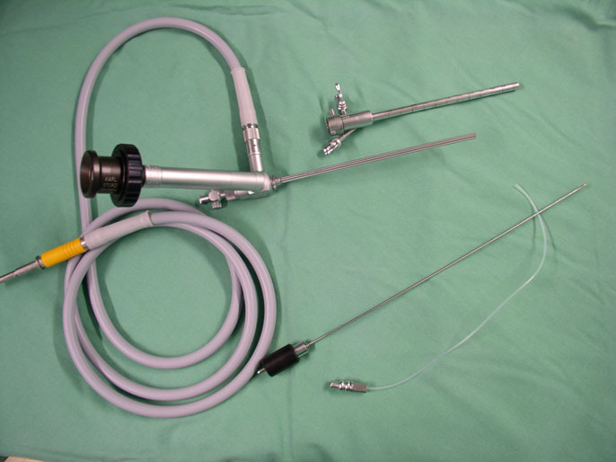 Neuroendoscopes
Using a neuro-endoscopic technique not only tumours or cysts located in the cavities of the braqin can be removed but also biopsies
of tumours or other lesions can be obtained under direct vision which considerably increases safety of the operation because intra-operative bleeding or other complications can be identified and treated immediately. It is also possible to use endoscopes to visualize structures which are normally difficult to reach. As it is possible to use angled optics the surgeon can virtually look around the corner and identify structures that otherwise could be not visualized and might be damaged. This is called endoscopy-assisted neurosurgery.
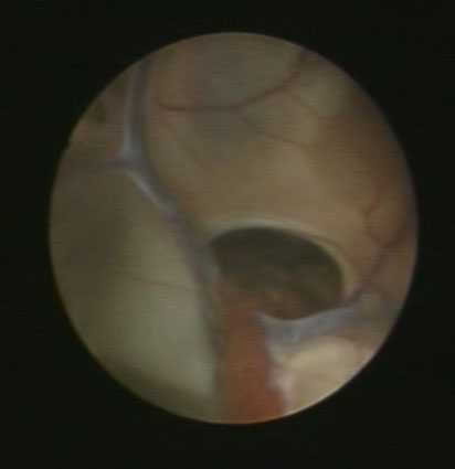
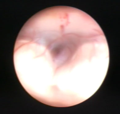
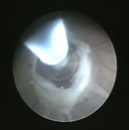
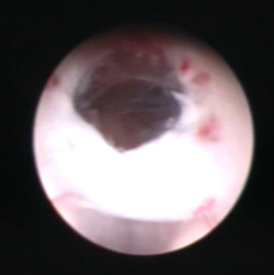
The most common neuro-endoscopic procedure is called endoscopic fenestration of the floor of the Third ventricle or "ETV". This method is used to treat certain forms of hydrocephalus - or commonly know as "water on the brain". Hydrocephalus means that the circulation of fluid in the brain is disrupted. This lead to high pressure in the patient's head which may give the patient headache, nausea and vomiting and even can make an emergency operation unavoidable. The most accepted and performed neruosurgical procedure to treat hydrocephalus is the implantation of a shunt.
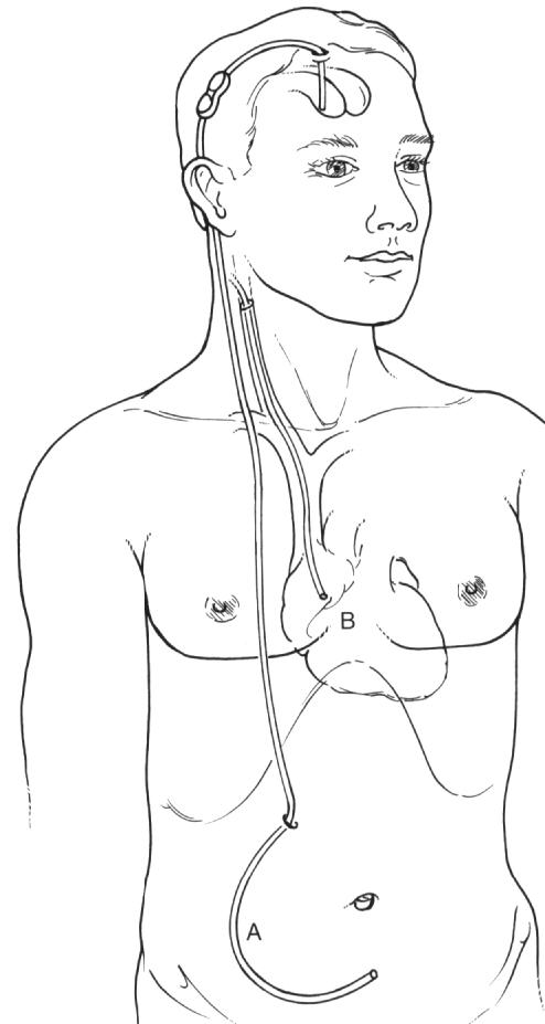
A shunt is a silicon tube which normally carries a small valve to regulate the flow of the CSF (cerebrospinal fluid). One end of the tube is inserted into the cavity of the brain. The fluid is diverted from the inside of the brain to a different part of the body where it can be absorbed. Therefore the other end of the shunt system is either placed in the peritoneal cavity or into the chest or even the heart.
It is very common that the patient needs the shunt for th rest of his or her life. So he needs to see a neurosurgeon on a more or less regular basis. Although the insertion of a shunt is a relatively easy procedure, there are many possible complications related to the shunt. As the patient carries the system for many years, infection, blockage of the system, over- or underdrainage are common problems.
There are certain types of hydrocephalus like a congenital narrowing of certain parts of the "pipe system" in the brain that can and should be treated
endoscopically. Lesions like colloid cysts (cysts which are filled with slime and block the CSF pathways) or tumours can be removed endoscopically with a rather small surgical risk. Very often, the patient is cured after the surgery and doesn't need further neurosurgical treatment. The complication rate is reduced to the direct risk of the procedure with no risk of long term complications.
 .
.  .
.  .
. 
Recently, there is a lot of development in the neuro-endoscopic field. New devices and techniques have been developed and more neurosurgical conditions can be treated endoscopically. I.e. it is sometimes possible to remove lumbar or cervical disc prolapses with an endoscopic technique, minimizing the surgicaltrauma substantially. Pituitary tumours are being removed routinely with an endoscopic technique through the nose of the patient. This is highly accepted by the patient as it also avoids an conventional opening of the skull and the trauma for the patient as well as time in hospital are remarkably reduced.
The use of endoscopic techniques in neurosurgery is substantially different to commonly used methods. The neurosurgeon should be trained accordingly and it is necessary to carry out neuro-endoscopic procedures on a regular basis in order to have enough experience and expertise. Major risks of neuro-endoscopic procedures are intra-operative bleeding (which can be very problematic due to the small approach) and post-operative infection. In unexperienced hands an endoscopic procedure can be very long which increases the complication rate.
In any case it is worthwhile to consider the possibility of an endoscopic neurosurgical procedure. Patients should consult a neurosurgeon who is capable of performing both neuro-endoscopic as well as conventional neurosurgical procedures to obtain an objective opinion.
Neuroendoscopy(![]() Deutsche Version)
Deutsche Version)
Please use the links in the header of this homepage if you would like to make arrangements for a confidential consultation !
J.A.Horaczek MD